Amazing new technology lets scientists watch fetuses develop in real time : ScienceAlert

To get a closer, real-time look at fetal development and better understand the potential causes of birth defects and other health problems, scientists have turned to a source you might not expect: quail eggs.
In fact, our earliest development as living beings is similar to that of quail, and because their embryos grow inside eggs, they can be scanned relatively easily. Bird eggs have long been favored by scientists for studying embryos.
Here, researchers in Australia used eggs carrying quail bred to express a fluorescent peptide that binds to actin proteins that form the structure of the early embryo, called the actin cytoskeleton. This approach allowed them to observe cells migrating and coming together to form organs.
“For the first time, we have seen high-resolution, real-time images of important early developmental processes,” says developmental biologist Melanie White from the University of Queensland.
“Until now, most of our knowledge about post-implantation development has come from studies on static slides, at fixed points in time.”
frameborder=”0″ allow=”accelerometer; autoplay; clipboard save; encrypted media; gyroscope; picture-in-picture; web sharing” referrerpolicy=”strict-origin-when-cross-origin” allowfullscreen>
The team was able to see the early stages of the formation of the heart, brain and spinal cord. A variety of microscope instruments were used to capture the fluorescent marker, which outlined the movement of the cells.
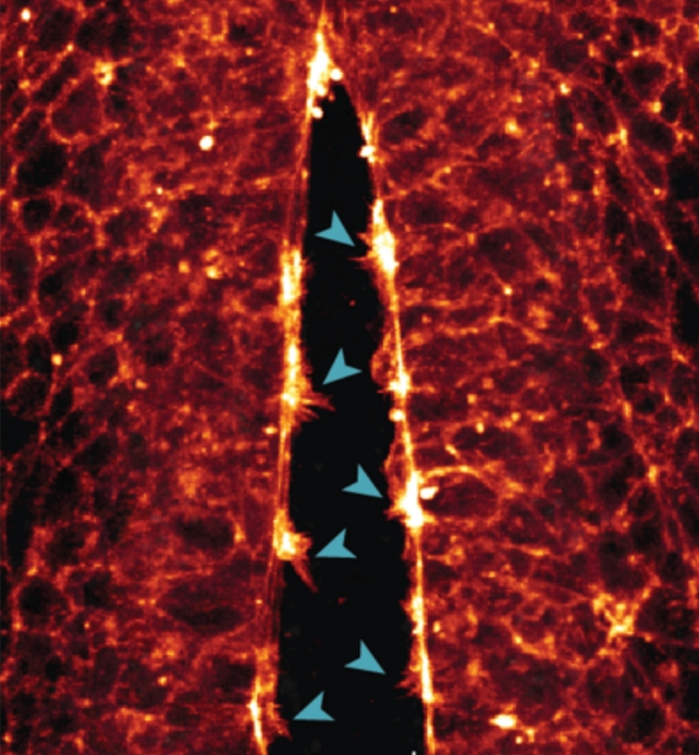
One of the observations made was of the neural tube, the precursor to the central nervous system, being “compressed” as the cells joined together.
“We saw how the cells crossed the open neural tube with their protrusions to contact the opposite side – the more protrusions the cells formed, the faster the tube closed,” explains White.
“If this process goes wrong or is interrupted and the tube does not close properly during the fourth week of human development, the embryo will have defects in the brain and spinal cord.”
Similar connections were made in the stem cells that would eventually form quail hearts.
“We were able to image the filopodia of heart stem cells inside the embryo as they first made contact, projecting protrusions and grasping at the environment and each other to form the early heart,” says White.
“This is the first time anyone has captured the cell’s actin cytoskeleton, facilitating this contact in live imaging.”
As well as offering a fascinating insight into early life, the study is important for increasing our knowledge of how and why birth defects occur. When the wiring processes fail, it can lead to problems for the developing baby.
Seeing these biological transformations happen in real time, and on the smallest scales, should be useful in the future for mitigating or at least identifying the risk of birth defects. Many more quail egg studies using this process are now planned by the team.
Scientists continue to improve their models and their understanding of what happens in the womb, and through this we can work to make more pregnancies as healthy as possible.
“Our goal is to find proteins or genes that could be targeted in the future or used to screen for birth defects,” says White.
“We are very excited about the possibilities this new quail model now offers for studying development in real time.”
The research was published in The Journal of Cell Biology.